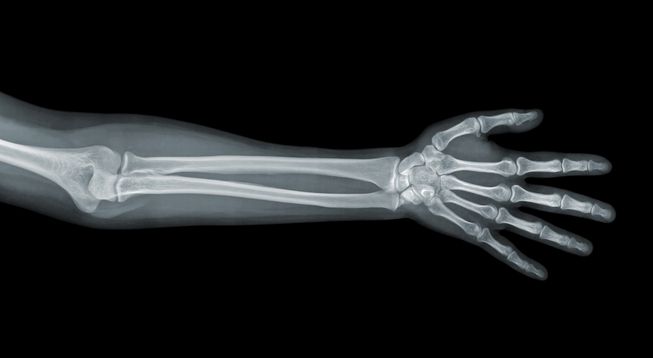
The discovery of X-ray has transformed medical diagnosis in human history. X-ray has provided doctors a way to understand internal physical situation of patients without having to perform a surgery. These X-ray images have always been black and white in the last century. Now, a groundbreaking element is introduced.
X-ray goes through our bodies. The varying densities in different parts absorb and reflect varying degrees of rays and beams. Softer, less dense tissues, such as skins, allow more rays to pass through whereas denser tissues, such as bones, absorb more beams and therefore, appear to be whiter. In short, the higher the density of our body parts, the lower the X-ray permeability, and thus, the whiter the images are, and vice versa.

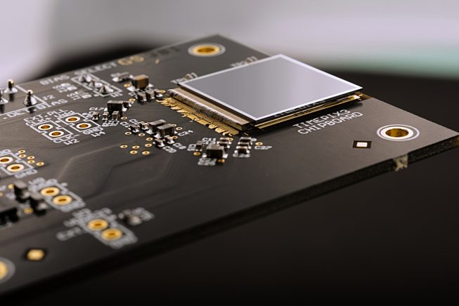
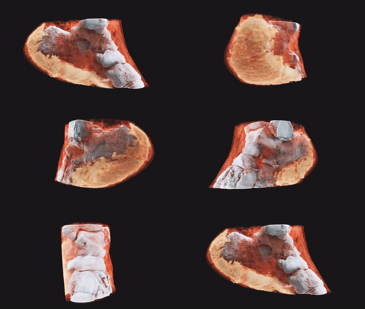
New Zealand company Mars Bioimaging has developed the newest version of medical scanning machine and completed the first human 3D colour X-ray! With the base of traditional X-ray machine and Higgs Boson particle found by European Organization for Nuclear Research (CERN), the system functions like a camera. At the moment the X-ray is shot, the Medipix3 particle tracking chip calculates the particles’ movement within the range, resulting in accurate X-ray images.

The particle tracking chip is able to detect the wavelength when X-rays permeate through our bodies and illustrate the difference between the body parts even more comprehensively, including not only bones and muscles but also fats, body fluids and so on. Additional softwares translate data into the colourful X-ray images, fostering doctors’ understanding of patients’ internals.

At present, such medical scanning machines are applicable in bones and joints checking, cardiovascular disease diagnosis, cancer research and there will only be more applications in the near future!


